
Histopathology and cytology analyzes
Reliable detection of neoplasia, determining whether the neoplasia is malignant or benign, detection of pathological changes of the eye that are allergic or autoimmune in nature, detection of the degree of the anomaly of the tissues and cells that build the eye, is possible by histopathology and cytology examination in our laboratory.
The owner may present a pet that is blind and has multiple pathological changes in the eye, and it is difficult to determine which pathological change was primary, and which changes occurred secondarily.
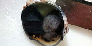
In these situations, we advise enucleation of the eye globe for diagnostic purposes, and also to solve the patient’s chronic pain and chronic source of inflammation. After enucleation, the eye globe is sent for histopathology examination which is of particular importance because it may save the healthy eye. This is because many eye diseases are bilateral, ie. they occur in both eyes, but the symptoms may not occur in both eyes at the same time. Also, eye tumors that are primary or metastatic often manifest clinically as inflammation of the eye and we are unable to see the obvious “mass” in the eye. These are just some of the reasons that teach us that:
EVERY EYE GLOBE SHOULD BE SENT TO HISTOPATHOLOGY AFTER ENUCLEATION!